Comprehensive Eye Exams
The visual symptoms of poor eye health can manifest themselves gradually, which is why many ophthalmologists and eye care professionals suggest annual eye exams. Changes can be so subtle they go unnoticed until vision is severely affected.
Regular comprehensive eye exams can detect eye disease as well as other conditions that may lead to vision loss if left untreated.
Eye exams may also give your Ocala Eye ophthalmologist clues to the health of other systems in the body.
What Happens During an Eye Exam?
 Several factors can help determine the extensiveness of the eye exam including:
- The patient’s medical history
- Any complaints about vision
- How long it has been since their last complete exam.
Before more detailed tests are performed, there are several initial evaluations that can give the eye doctor some clues as to how one’s overall vision is functioning. Usually, this starts with the autorefractor, a device used to check for refractive errors. One eye is tested at a time using a series of pictures.
After the autorefractor test, evaluation of the visual fields, pupils, and extraocular muscle function are performed.
Once this is done, the more detailed portion of the eye exam usually follows a “front to back” analysis of the eye, beginning with the refraction. While sitting in the exam chair, patients will be asked to identify letters on a wall chart to help determine visual acuity. An instrument called a phoropter is then placed in front the eyes to determine if vision can be improved with eyeglasses. The phoropter houses a multitude of lenses that can be easily rotated to help the clinician determine a prescription.
Intraocular Pressure
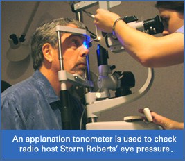
 Ocala Eye uses what’s known as an applanation tonometer to determine the internal pressure of the eye. High intraocular pressure is an indicator of the potential for developing glaucoma. Numbing eye drops are administered along with fluorescein dye prior to using the tonometer. A blue light is used to assist in the intraocular pressure measurement as well as determining irregularities on the corneal surface.
Dilated Exam
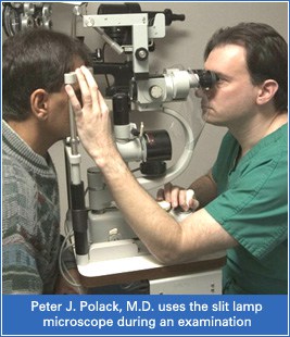
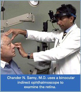
With the initial data gathered from the aforementioned tests, the Ocala Eye ophthalmologist or optometrist will need to more closely examine the structures of the eye. Examination takes place using a slit-lamp microscope and a head-mounted binocular indirect ophthalmoscope (BIO). This part of the examination requires the pupils to be dilated. Eye drops are administered to relax your iris and increase pupil diameter.
The slit lamp microscope has a support where patients rest their chin and stabilize their head. The eye doctor then uses a narrow beam of different wavelengths (colors) of light to show details specific to various eye structures.
 The eye doctor first examines the eyes in layers using the slit lamp microscope, starting with the anterior (front) structures like the cornea, eyelids and tear ducts then progresses further into the eye. Middle structures like the lens and vitreous are examined next followed by the posterior (back) structures including the retina and optic nerve.
After the slit lamp evaluation, your doctor will continue with the examination using the binocular indirect ophthalmoscope. When combined with a hand-held lens, the BIO allows the doctor to see peripheral features of the retina.
When the examination is complete, your Ocala Eye doctor will discuss their findings and offer a top-level overall description of their findings. With many eye diseases, early detection can have a significant effect on treatment options.
Remember, regular eye exams help eye doctors gather important information that can help to preserve vision for a lifetime.
Consult an Ophthalmologist
Ocala Eye’s fellowship-trained ophthalmologists and medical staff are dedicated to helping your vision last a lifetime, which is why we offer comprehensive eye care for adults of all ages. From annual eye exams to advanced procedures like LASIK and cataract surgery, we can help you see your best at any phase of your life.
